Microscopy and Microorganisms
Microscopes
Microscopes are useful for showing detail at cellular and sub-cellular level and are used to observe objects that are too small to see with the naked eye.
There are two main types of microscope – the Light Microscope (like the ones you will use in your school or college) and the Electron Microscope which has higher magnifications and resolution than a Light Microscope (used by scientists).
The Light Microscope
Image
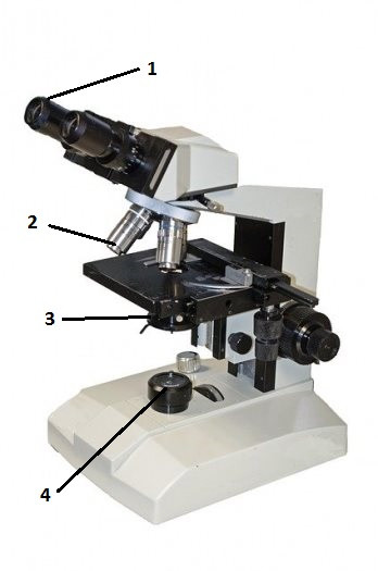
1 The Eye piece
2 Objective lenses of different magnifications
3 The Iris
3 The Light source.
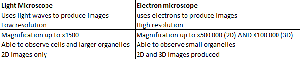
Comparing light and electron microscopes
Image
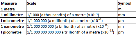
Common units used in microscopy
Image
Magnification and resolution
Magnification is the measure of how many times a given object under a microscope has been made larger.
To calculate the magnifying power of a microscope you need to use the below formula:
Magnification = size of the image / size of the real object
An example question you may be asked could be: A light microscope produces an image of a cell which has a diameter of 1600µm. The cell’s actual diameter is 40µm. Calculate the magnifying power of a microscope.
Magnification = 1600/40 = 40
Resolution
Resolution is the smallest distance between two points on a specimen that can still be identified. When looking through a light microscope you can see the fine detail down to 200 nanometres. Electron microscopes have the power to see detail down to 0.05 nanometres.
Organelles can be made more visible on a microscope by using staining techniques.
Methylene blue is often used to stain the nuclei of animal cells for viewing under a light microscope. Heavy metals which include cadmium are often used to stain specimens for viewing under a powerful electron microscope.
Culturing microorganisms
To aid research uncontaminated samples are cultured or grown in the laboratory.
In order to grow microorganisms need the following:
· A source of food – usually a nutrient and or an agar.
· A warm environment – in schools this is typically 25⁰c as it lowers the possibility of pathogens growing
· A source of moisture
Aseptic technique
When culturing microorganisms in the laboratory aseptic technique is used to:
· Produce uncontaminated cultures
· Reduce the risk of infection to scientists by pathogens
Bacterial growth
A bacterial cell will grow by binary fission, usually in a few days. In this process a cell will usually divide in around 20 minutes. The bacteria will continue to divide in this way until the numbers are in their millions. As they grow cells will form circular colonies which will become visible to the naked eye. To estimate their growth rate you can count them or calculate their area.
Category